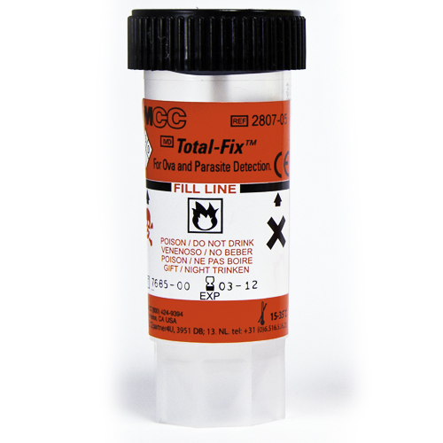
Species of microsporidia have been associated with severe infections in AIDS patients. Encephalocytozoon hellum (keratoconjunctivitas), E. cuniculi (hepatitis 7 peritonitis), and Enterocytozoon bieneusi (diarrhea) are among the most commonly reported. In 1992, Weber et al. published a modified trichrome staining procedure for microsporidial spores in duodenal aspirates. A slightly modified procedure was recommended by the CDC. In 1993, Ryan et al. described a trichrome-blue modification of the Weber et al. procedure. The trichrome-blue staining procedure can be used to detect microsporidial species in urine, stool, and nasopharyngeal specimens. Spores stain pink-red with a polar or central clear zone, whereas yeast cells and pseudohyphae stain a grayish blue color.
PRODUCT # |
DESCRIPTION |
601A | Ryan modification of Trichrome-Blue |
3719A | 95% Reagent Alcohol |
374B | Reagent Alcohol |
3720A | Trichrome Decolorizer |
134B | Xylene |
930E | Xylene Substitute (d-limonene) |
Thin smears should be made from formalin preserved samples, collected in the usual manner. Smears should be air dried and fixed in absolute alcohol for 5 minutes prior to staining.
*an acceptable xylene-substitute can be used
Note: Staining times may vary to suit the individual.
Preparations should be read at 1000x magnification. At least 100 fields should be read before reporting a negative result. Microsporidia are oval, and measure 0.7-1.1 x 1.1-1.7 µm. The spore wall stains a bright pinkish-red. The spore is transparent or will exhibit a pinkish belt-like stripe across the center.